
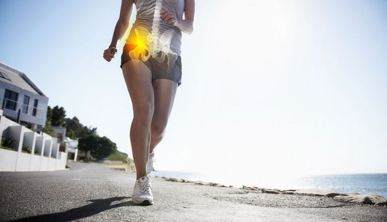
Who hasn’t heard—or even uttered—a phrase like this? But unlike other, more localized areas of our body, joints such as the knee or ankle; or muscles like the calf, hamstrings, or quadriceps, the hip is far more involved, and in that spot of our anatomical world many other things happen. Here we help you identify them.
We tend to call the hip anything in the groin area and the outer side of the thigh, but in reality the hip is only the joint between the femur and the pelvis, and it sits “within fingertip reach” in the middle of the groin line. And thankfully not everything that hurts there is the hip.
This year Killian finished the Eiger Ultra in 46th place, according to reports afterward; he began to notice some discomfort in his left hip, and what initially seemed like a strain or tendinopathy turned out to be a bone edema in the hip. Later he attributed it to not having made a proper transition back into training after his stay in the Himalayas and neglecting his strength work. Probably he wasn’t far off.
This year I’ve treated several patients with similar hip and pelvis injuries.
What causes this injury and what does it involve?
The hip is a special joint; anatomically it is very similar to the shoulder (glenohumeral joint) but differs in function. Just as the shoulder is a mobility-focused joint and the arm bone articulates “straight” in the glenoid, in the case of the hip it is a weight-bearing joint, and because of upright standing, the femur acts like a rod with the femoral head at the end of the “handle” and the muscular insertions that rotate the femur on the other side of that handle, at the trochanter.
Being a weight-bearing joint, there is a critical area at the femoral neck (the region between the trochanter—the muscle insertion zone on the lateral thigh—and the articular head). That area bears the load of the limb, or to put it more precisely, endures the load like the eye of a bridge between the femur and the joint. It is the region where most fractures occur.
The joint surface, the head, can suffer varying degrees of injury due to that load, ranging from edema to cartilage wear or labral tears (the hip’s meniscus).
These injuries arise from excessive loading of the joint: overly aggressive training with a lot of impact, too many competitions, weakness of the muscles that must stabilize the joint…
The bone edema I mentioned a while back is simply what shows up on an MRI; it reflects microruptures within the bone’s inner trabeculae, translating into bleeding and internal edema.
The problem is that edema in a muscle or other soft tissues (subcutaneous tissue, etc.) generally resolves in a few weeks. Bone edema, which tends to appear in load-bearing zones, does not resolve as quickly (it can take months), and its treatment, as we explained before, is challenging (rest and magnetotherapy are part of the fundamental treatment). Therefore, you must switch activities and “enjoy” cycling, swimming, and moderate walks during the recovery period.
Another group of injuries related to this hip pain are tendinopathies and bursitis; with trochanteritis (pain on the outer side of the thigh) and pubalgia (pain at the adductor insertions on the pubis) being the most common. These are mainly overuse injuries (trochanteritis can also occur from blows), due to insufficient strengthening work or a sharp increase in training or competition load. Not everything is running.
How should it be treated?
Treatment for these injuries involves physical therapy across its various techniques, and even corticosteroid injections, growth factors, and other therapeutic methods.
Pain in the hip does not always originate from the joint or the working muscles; sometimes the source is injuries to the lumbar nerves that affect the region and trigger referred pain.
Another frequent origin is inguinal hernias, which can produce pain in the area and are often solved surgically.
So you see, that “my hip hurts” isn’t always straightforward, and it isn’t always the hip; when a patient presents with that complaint, I always think of the phrase “my kidneys hurt” as a reference to lumbar pain.
Therefore, listen to the athlete, investigate the pain’s cause (changes in training terrain, excessive increases in load, footwear changes…), perform a proper examination, and order the appropriate imaging tests (X-rays, ultrasounds, MRIs, CT scans…), always with a clear orientation and knowing what we are trying to look for.
From there, implement the most appropriate treatment and be patient, respecting the rehabilitation timeline, and not getting fixated on maintaining the usual activity at all costs.
